Figure 4
3D software reconstruction for planning robotic assisted radical nephrectomy with level III caval thrombus
Marcos Tobias-Machado*, Ricardo JF de Bragança, Rafael Tourinho-Barbosa, Hamilton C Zampolli and Aurus M Dourado
Published: 30 April, 2020 | Volume 4 - Issue 1 | Pages: 029-033
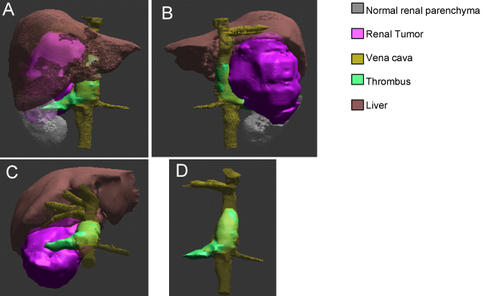
Figure 4:
3D holographic reconstruction: A. anterior view showing perfectly the relation between renal tumor, normal renal parenchyma, liver, veins (renal, hepatic and vena cava) and caval thombus. B. Posterior view showing better the renal tumo and caval thombus. C. topographic anterior view. D. subtracted image showing thrombus in the right renal vein and vena cava up close to the hepatic veins.
Read Full Article HTML DOI: 10.29328/journal.acst.1001019 Cite this Article Read Full Article PDF
More Images
Similar Articles
-
Theranostics: A Unique Concept to Nuclear MedicineLucio Mango*. Theranostics: A Unique Concept to Nuclear Medicine. . 2017 doi: 10.29328/journal.acst.1001001; 1: 001-004
-
Predictors of Candidemia infections and its associated risk of mortality among adult and pediatric cancer patients: A retrospective study in Lahore, Punjab, PakistanHafiz Muhammad Bilal*,Neelam Iqbal,Shazia Ayaz. Predictors of Candidemia infections and its associated risk of mortality among adult and pediatric cancer patients: A retrospective study in Lahore, Punjab, Pakistan. . 2018 doi: 10.29328/journal.acst.1001003; 2: 001-007
-
Endogenus toxicology: Modern physio-pathological aspects and relationship with new therapeutic strategies. An integrative discipline incorporating concepts from different research discipline like Biochemistry, Pharmacology and ToxicologyLuisetto M*,Naseer Almukhtar,Behzad Nili Ahmadabadi,Gamal Abdul Hamid,Ghulam Rasool Mashori,Kausar Rehman Khan,Farhan Ahmad Khan,Luca Cabianca. Endogenus toxicology: Modern physio-pathological aspects and relationship with new therapeutic strategies. An integrative discipline incorporating concepts from different research discipline like Biochemistry, Pharmacology and Toxicology . . 2019 doi: 10.29328/journal.acst.1001004; 3: 001-024
-
Insilico investigation of TNFSF10 signaling cascade in ovarian serous cystadenocarcinomaAsima Tayyeb*,Zafar Abbas Shah. Insilico investigation of TNFSF10 signaling cascade in ovarian serous cystadenocarcinoma. . 2019 doi: 10.29328/journal.acst.1001005; 3: 025-034
-
Fifth “dark” force completely change our understanding of the universeRobert Skopec*. Fifth “dark” force completely change our understanding of the universe. . 2019 doi: 10.29328/journal.acst.1001006; 3: 035-041
-
Risk factors of survival in breast cancerAkram Yazdani*. Risk factors of survival in breast cancer. . 2019 doi: 10.29328/journal.acst.1001007; 3: 042-044
-
Results of chemotherapy in the treatment of chronic lymphoid leukemia in Black Africa: Experience of Côte d’IvoirePacko Dieu-le-veut Saint-Cyr Sylvestre*,N’dhatz Comoe Emeraude,Kamara Ismael,Boidy Kouakou,Koffi Kouassi Gustave,Nanho Danho Clotaire,Koffi Kouassi Gustave. Results of chemotherapy in the treatment of chronic lymphoid leukemia in Black Africa: Experience of Côte d’Ivoire. . 2019 doi: 10.29328/journal.acst.1001008; 3: 045-048
-
Stercoral perforation: A rare case and reviewLava Krishna Kannappa*,Muhammad Sufian Khalid,May Hnin Lwin Ko,Mohsin Hussein,Jia Hui Choong,Ameer Omar Rawal-Pangarkar,Danaradja Armugam,Yahya Salama. Stercoral perforation: A rare case and review. . 2019 doi: 10.29328/journal.acst.1001009; 3: 049-051
-
Different optimization strategies for the optimal control of tumor growthAbd El Moniem NK*,Sweilam NH,Tharwat AA. Different optimization strategies for the optimal control of tumor growth. . 2019 doi: 10.29328/journal.acst.1001010; 3: 052-062
-
Risk factor of liver metastases in breast cancerAkram Yazdani*. Risk factor of liver metastases in breast cancer. . 2019 doi: 10.29328/journal.acst.1001011; 3: 063-065
Recently Viewed
-
Cancer Cell Resistance: The Emergent Intelligence of Adaptation and the Need for Biophysical IntegrationMohamed H Doweidar*. Cancer Cell Resistance: The Emergent Intelligence of Adaptation and the Need for Biophysical Integration. Int J Clin Microbiol Biochem Technol. 2025: doi: 10.29328/journal.ijcmbt.1001031; 8: 007-008
-
Insilico investigation of TNFSF10 signaling cascade in ovarian serous cystadenocarcinomaAsima Tayyeb*,Zafar Abbas Shah. Insilico investigation of TNFSF10 signaling cascade in ovarian serous cystadenocarcinoma. Arch Cancer Sci Ther. 2019: doi: 10.29328/journal.acst.1001005; 3: 025-034
-
Why? It is the question that I ask myself every day, every hour, every second...Francisco Javier Torres-Gómez*. Why? It is the question that I ask myself every day, every hour, every second.... Arch Cancer Sci Ther. 2021: doi: 10.29328/journal.acst.1001024; 5: 006-006
-
Breast Cancer in FemaleLorena Menditto*. Breast Cancer in Female. Arch Cancer Sci Ther. 2024: doi: 10.29328/journal.acst.1001040; 8: 013-018
-
Heart failure with preserved ejection fraction: A mini reviewNathan Buila,Jean-René M’Buyamba-Kabangu*. Heart failure with preserved ejection fraction: A mini review. Arch Vas Med. 2022: doi: 10.29328/journal.avm.1001016; 6: 005-006
Most Viewed
-
Causal Link between Human Blood Metabolites and Asthma: An Investigation Using Mendelian RandomizationYong-Qing Zhu, Xiao-Yan Meng, Jing-Hua Yang*. Causal Link between Human Blood Metabolites and Asthma: An Investigation Using Mendelian Randomization. Arch Asthma Allergy Immunol. 2023 doi: 10.29328/journal.aaai.1001032; 7: 012-022
-
Impact of Latex Sensitization on Asthma and Rhinitis Progression: A Study at Abidjan-Cocody University Hospital - Côte d’Ivoire (Progression of Asthma and Rhinitis related to Latex Sensitization)Dasse Sery Romuald*, KL Siransy, N Koffi, RO Yeboah, EK Nguessan, HA Adou, VP Goran-Kouacou, AU Assi, JY Seri, S Moussa, D Oura, CL Memel, H Koya, E Atoukoula. Impact of Latex Sensitization on Asthma and Rhinitis Progression: A Study at Abidjan-Cocody University Hospital - Côte d’Ivoire (Progression of Asthma and Rhinitis related to Latex Sensitization). Arch Asthma Allergy Immunol. 2024 doi: 10.29328/journal.aaai.1001035; 8: 007-012
-
An algorithm to safely manage oral food challenge in an office-based setting for children with multiple food allergiesNathalie Cottel,Aïcha Dieme,Véronique Orcel,Yannick Chantran,Mélisande Bourgoin-Heck,Jocelyne Just. An algorithm to safely manage oral food challenge in an office-based setting for children with multiple food allergies. Arch Asthma Allergy Immunol. 2021 doi: 10.29328/journal.aaai.1001027; 5: 030-037
-
Snow white: an allergic girl?Oreste Vittore Brenna*. Snow white: an allergic girl?. Arch Asthma Allergy Immunol. 2022 doi: 10.29328/journal.aaai.1001029; 6: 001-002
-
Cytokine intoxication as a model of cell apoptosis and predict of schizophrenia - like affective disordersElena Viktorovna Drozdova*. Cytokine intoxication as a model of cell apoptosis and predict of schizophrenia - like affective disorders. Arch Asthma Allergy Immunol. 2021 doi: 10.29328/journal.aaai.1001028; 5: 038-040

If you are already a member of our network and need to keep track of any developments regarding a question you have already submitted, click "take me to my Query."


