Figure 3
3D software reconstruction for planning robotic assisted radical nephrectomy with level III caval thrombus
Marcos Tobias-Machado*, Ricardo JF de Bragança, Rafael Tourinho-Barbosa, Hamilton C Zampolli and Aurus M Dourado
Published: 30 April, 2020 | Volume 4 - Issue 1 | Pages: 029-033

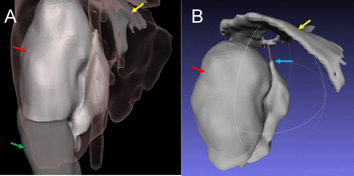
Figure 3:
3D holographic reconstruction: A. Anterior view showing perfectly the relation between renal tumor (red arrow), normal renal parenchyma (green arrow), hepatic border + vena cava (pink shadow) and diafragma (yellow arrow). B. Posterior view showing renal tumor (red arrow), distal extension of thrombus (blue arow) and diaphragm (yellow arrow). There are 3 cm above the thrombus to put a vessel loop to proximal control.
Read Full Article HTML DOI: 10.29328/journal.acst.1001019 Cite this Article Read Full Article PDF
More Images
Similar Articles
-
Theranostics: A Unique Concept to Nuclear MedicineLucio Mango*. Theranostics: A Unique Concept to Nuclear Medicine. . 2017 doi: 10.29328/journal.acst.1001001; 1: 001-004
-
Predictors of Candidemia infections and its associated risk of mortality among adult and pediatric cancer patients: A retrospective study in Lahore, Punjab, PakistanHafiz Muhammad Bilal*,Neelam Iqbal,Shazia Ayaz. Predictors of Candidemia infections and its associated risk of mortality among adult and pediatric cancer patients: A retrospective study in Lahore, Punjab, Pakistan. . 2018 doi: 10.29328/journal.acst.1001003; 2: 001-007
-
Endogenus toxicology: Modern physio-pathological aspects and relationship with new therapeutic strategies. An integrative discipline incorporating concepts from different research discipline like Biochemistry, Pharmacology and ToxicologyLuisetto M*,Naseer Almukhtar,Behzad Nili Ahmadabadi,Gamal Abdul Hamid,Ghulam Rasool Mashori,Kausar Rehman Khan,Farhan Ahmad Khan,Luca Cabianca. Endogenus toxicology: Modern physio-pathological aspects and relationship with new therapeutic strategies. An integrative discipline incorporating concepts from different research discipline like Biochemistry, Pharmacology and Toxicology . . 2019 doi: 10.29328/journal.acst.1001004; 3: 001-024
-
Insilico investigation of TNFSF10 signaling cascade in ovarian serous cystadenocarcinomaAsima Tayyeb*,Zafar Abbas Shah. Insilico investigation of TNFSF10 signaling cascade in ovarian serous cystadenocarcinoma. . 2019 doi: 10.29328/journal.acst.1001005; 3: 025-034
-
Fifth “dark” force completely change our understanding of the universeRobert Skopec*. Fifth “dark” force completely change our understanding of the universe. . 2019 doi: 10.29328/journal.acst.1001006; 3: 035-041
-
Risk factors of survival in breast cancerAkram Yazdani*. Risk factors of survival in breast cancer. . 2019 doi: 10.29328/journal.acst.1001007; 3: 042-044
-
Results of chemotherapy in the treatment of chronic lymphoid leukemia in Black Africa: Experience of Côte d’IvoirePacko Dieu-le-veut Saint-Cyr Sylvestre*,N’dhatz Comoe Emeraude,Kamara Ismael,Boidy Kouakou,Koffi Kouassi Gustave,Nanho Danho Clotaire,Koffi Kouassi Gustave. Results of chemotherapy in the treatment of chronic lymphoid leukemia in Black Africa: Experience of Côte d’Ivoire. . 2019 doi: 10.29328/journal.acst.1001008; 3: 045-048
-
Stercoral perforation: A rare case and reviewLava Krishna Kannappa*,Muhammad Sufian Khalid,May Hnin Lwin Ko,Mohsin Hussein,Jia Hui Choong,Ameer Omar Rawal-Pangarkar,Danaradja Armugam,Yahya Salama. Stercoral perforation: A rare case and review. . 2019 doi: 10.29328/journal.acst.1001009; 3: 049-051
-
Different optimization strategies for the optimal control of tumor growthAbd El Moniem NK*,Sweilam NH,Tharwat AA. Different optimization strategies for the optimal control of tumor growth. . 2019 doi: 10.29328/journal.acst.1001010; 3: 052-062
-
Risk factor of liver metastases in breast cancerAkram Yazdani*. Risk factor of liver metastases in breast cancer. . 2019 doi: 10.29328/journal.acst.1001011; 3: 063-065
Recently Viewed
-
Sensitivity and Intertextile variance of amylase paper for saliva detectionAlexander Lotozynski*. Sensitivity and Intertextile variance of amylase paper for saliva detection. J Forensic Sci Res. 2020: doi: 10.29328/journal.jfsr.1001017; 4: 001-003
-
Extraction of DNA from face mask recovered from a kidnapping sceneBassey Nsor*,Inuwa HM. Extraction of DNA from face mask recovered from a kidnapping scene. J Forensic Sci Res. 2022: doi: 10.29328/journal.jfsr.1001029; 6: 001-005
-
The Ketogenic Diet: The Ke(y) - to Success? A Review of Weight Loss, Lipids, and Cardiovascular RiskAngela H Boal*, Christina Kanonidou. The Ketogenic Diet: The Ke(y) - to Success? A Review of Weight Loss, Lipids, and Cardiovascular Risk. J Cardiol Cardiovasc Med. 2024: doi: 10.29328/journal.jccm.1001178; 9: 052-057
-
Could apple cider vinegar be used for health improvement and weight loss?Alexander V Sirotkin*. Could apple cider vinegar be used for health improvement and weight loss?. New Insights Obes Gene Beyond. 2021: doi: 10.29328/journal.niogb.1001016; 5: 014-016
-
Maximizing the Potential of Ketogenic Dieting as a Potent, Safe, Easy-to-Apply and Cost-Effective Anti-Cancer TherapySimeon Ikechukwu Egba*,Daniel Chigbo. Maximizing the Potential of Ketogenic Dieting as a Potent, Safe, Easy-to-Apply and Cost-Effective Anti-Cancer Therapy. Arch Cancer Sci Ther. 2025: doi: 10.29328/journal.acst.1001047; 9: 001-005
Most Viewed
-
Evaluation of Biostimulants Based on Recovered Protein Hydrolysates from Animal By-products as Plant Growth EnhancersH Pérez-Aguilar*, M Lacruz-Asaro, F Arán-Ais. Evaluation of Biostimulants Based on Recovered Protein Hydrolysates from Animal By-products as Plant Growth Enhancers. J Plant Sci Phytopathol. 2023 doi: 10.29328/journal.jpsp.1001104; 7: 042-047
-
Sinonasal Myxoma Extending into the Orbit in a 4-Year Old: A Case PresentationJulian A Purrinos*, Ramzi Younis. Sinonasal Myxoma Extending into the Orbit in a 4-Year Old: A Case Presentation. Arch Case Rep. 2024 doi: 10.29328/journal.acr.1001099; 8: 075-077
-
Feasibility study of magnetic sensing for detecting single-neuron action potentialsDenis Tonini,Kai Wu,Renata Saha,Jian-Ping Wang*. Feasibility study of magnetic sensing for detecting single-neuron action potentials. Ann Biomed Sci Eng. 2022 doi: 10.29328/journal.abse.1001018; 6: 019-029
-
Pediatric Dysgerminoma: Unveiling a Rare Ovarian TumorFaten Limaiem*, Khalil Saffar, Ahmed Halouani. Pediatric Dysgerminoma: Unveiling a Rare Ovarian Tumor. Arch Case Rep. 2024 doi: 10.29328/journal.acr.1001087; 8: 010-013
-
Physical activity can change the physiological and psychological circumstances during COVID-19 pandemic: A narrative reviewKhashayar Maroufi*. Physical activity can change the physiological and psychological circumstances during COVID-19 pandemic: A narrative review. J Sports Med Ther. 2021 doi: 10.29328/journal.jsmt.1001051; 6: 001-007

HSPI: We're glad you're here. Please click "create a new Query" if you are a new visitor to our website and need further information from us.
If you are already a member of our network and need to keep track of any developments regarding a question you have already submitted, click "take me to my Query."


