Figure 1
3D software reconstruction for planning robotic assisted radical nephrectomy with level III caval thrombus
Marcos Tobias-Machado*, Ricardo JF de Bragança, Rafael Tourinho-Barbosa, Hamilton C Zampolli and Aurus M Dourado
Published: 30 April, 2020 | Volume 4 - Issue 1 | Pages: 029-033

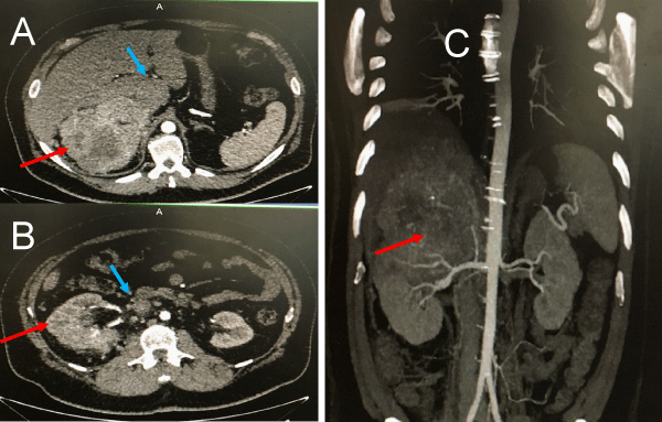
Figure 1:
CT scan images (A and B. coronal, C.Sagital) showing a large tumor mass (red arrow) with caval thrombus (blue arrow) identified until infra hepatic region. In this image is not possible to know the exact level of distal extension of caval thrombus.
Read Full Article HTML DOI: 10.29328/journal.acst.1001019 Cite this Article Read Full Article PDF
More Images
Similar Articles
-
Theranostics: A Unique Concept to Nuclear MedicineLucio Mango*. Theranostics: A Unique Concept to Nuclear Medicine. . 2017 doi: 10.29328/journal.acst.1001001; 1: 001-004
-
Predictors of Candidemia infections and its associated risk of mortality among adult and pediatric cancer patients: A retrospective study in Lahore, Punjab, PakistanHafiz Muhammad Bilal*,Neelam Iqbal,Shazia Ayaz. Predictors of Candidemia infections and its associated risk of mortality among adult and pediatric cancer patients: A retrospective study in Lahore, Punjab, Pakistan. . 2018 doi: 10.29328/journal.acst.1001003; 2: 001-007
-
Endogenus toxicology: Modern physio-pathological aspects and relationship with new therapeutic strategies. An integrative discipline incorporating concepts from different research discipline like Biochemistry, Pharmacology and ToxicologyLuisetto M*,Naseer Almukhtar,Behzad Nili Ahmadabadi,Gamal Abdul Hamid,Ghulam Rasool Mashori,Kausar Rehman Khan,Farhan Ahmad Khan,Luca Cabianca. Endogenus toxicology: Modern physio-pathological aspects and relationship with new therapeutic strategies. An integrative discipline incorporating concepts from different research discipline like Biochemistry, Pharmacology and Toxicology . . 2019 doi: 10.29328/journal.acst.1001004; 3: 001-024
-
Insilico investigation of TNFSF10 signaling cascade in ovarian serous cystadenocarcinomaAsima Tayyeb*,Zafar Abbas Shah. Insilico investigation of TNFSF10 signaling cascade in ovarian serous cystadenocarcinoma. . 2019 doi: 10.29328/journal.acst.1001005; 3: 025-034
-
Fifth “dark” force completely change our understanding of the universeRobert Skopec*. Fifth “dark” force completely change our understanding of the universe. . 2019 doi: 10.29328/journal.acst.1001006; 3: 035-041
-
Risk factors of survival in breast cancerAkram Yazdani*. Risk factors of survival in breast cancer. . 2019 doi: 10.29328/journal.acst.1001007; 3: 042-044
-
Results of chemotherapy in the treatment of chronic lymphoid leukemia in Black Africa: Experience of Côte d’IvoirePacko Dieu-le-veut Saint-Cyr Sylvestre*,N’dhatz Comoe Emeraude,Kamara Ismael,Boidy Kouakou,Koffi Kouassi Gustave,Nanho Danho Clotaire,Koffi Kouassi Gustave. Results of chemotherapy in the treatment of chronic lymphoid leukemia in Black Africa: Experience of Côte d’Ivoire. . 2019 doi: 10.29328/journal.acst.1001008; 3: 045-048
-
Stercoral perforation: A rare case and reviewLava Krishna Kannappa*,Muhammad Sufian Khalid,May Hnin Lwin Ko,Mohsin Hussein,Jia Hui Choong,Ameer Omar Rawal-Pangarkar,Danaradja Armugam,Yahya Salama. Stercoral perforation: A rare case and review. . 2019 doi: 10.29328/journal.acst.1001009; 3: 049-051
-
Different optimization strategies for the optimal control of tumor growthAbd El Moniem NK*,Sweilam NH,Tharwat AA. Different optimization strategies for the optimal control of tumor growth. . 2019 doi: 10.29328/journal.acst.1001010; 3: 052-062
-
Risk factor of liver metastases in breast cancerAkram Yazdani*. Risk factor of liver metastases in breast cancer. . 2019 doi: 10.29328/journal.acst.1001011; 3: 063-065
Recently Viewed
-
Sensitivity and Intertextile variance of amylase paper for saliva detectionAlexander Lotozynski*. Sensitivity and Intertextile variance of amylase paper for saliva detection. J Forensic Sci Res. 2020: doi: 10.29328/journal.jfsr.1001017; 4: 001-003
-
Extraction of DNA from face mask recovered from a kidnapping sceneBassey Nsor*,Inuwa HM. Extraction of DNA from face mask recovered from a kidnapping scene. J Forensic Sci Res. 2022: doi: 10.29328/journal.jfsr.1001029; 6: 001-005
-
The Ketogenic Diet: The Ke(y) - to Success? A Review of Weight Loss, Lipids, and Cardiovascular RiskAngela H Boal*, Christina Kanonidou. The Ketogenic Diet: The Ke(y) - to Success? A Review of Weight Loss, Lipids, and Cardiovascular Risk. J Cardiol Cardiovasc Med. 2024: doi: 10.29328/journal.jccm.1001178; 9: 052-057
-
Could apple cider vinegar be used for health improvement and weight loss?Alexander V Sirotkin*. Could apple cider vinegar be used for health improvement and weight loss?. New Insights Obes Gene Beyond. 2021: doi: 10.29328/journal.niogb.1001016; 5: 014-016
-
Maximizing the Potential of Ketogenic Dieting as a Potent, Safe, Easy-to-Apply and Cost-Effective Anti-Cancer TherapySimeon Ikechukwu Egba*,Daniel Chigbo. Maximizing the Potential of Ketogenic Dieting as a Potent, Safe, Easy-to-Apply and Cost-Effective Anti-Cancer Therapy. Arch Cancer Sci Ther. 2025: doi: 10.29328/journal.acst.1001047; 9: 001-005
Most Viewed
-
Evaluation of Biostimulants Based on Recovered Protein Hydrolysates from Animal By-products as Plant Growth EnhancersH Pérez-Aguilar*, M Lacruz-Asaro, F Arán-Ais. Evaluation of Biostimulants Based on Recovered Protein Hydrolysates from Animal By-products as Plant Growth Enhancers. J Plant Sci Phytopathol. 2023 doi: 10.29328/journal.jpsp.1001104; 7: 042-047
-
Sinonasal Myxoma Extending into the Orbit in a 4-Year Old: A Case PresentationJulian A Purrinos*, Ramzi Younis. Sinonasal Myxoma Extending into the Orbit in a 4-Year Old: A Case Presentation. Arch Case Rep. 2024 doi: 10.29328/journal.acr.1001099; 8: 075-077
-
Feasibility study of magnetic sensing for detecting single-neuron action potentialsDenis Tonini,Kai Wu,Renata Saha,Jian-Ping Wang*. Feasibility study of magnetic sensing for detecting single-neuron action potentials. Ann Biomed Sci Eng. 2022 doi: 10.29328/journal.abse.1001018; 6: 019-029
-
Pediatric Dysgerminoma: Unveiling a Rare Ovarian TumorFaten Limaiem*, Khalil Saffar, Ahmed Halouani. Pediatric Dysgerminoma: Unveiling a Rare Ovarian Tumor. Arch Case Rep. 2024 doi: 10.29328/journal.acr.1001087; 8: 010-013
-
Physical activity can change the physiological and psychological circumstances during COVID-19 pandemic: A narrative reviewKhashayar Maroufi*. Physical activity can change the physiological and psychological circumstances during COVID-19 pandemic: A narrative review. J Sports Med Ther. 2021 doi: 10.29328/journal.jsmt.1001051; 6: 001-007

HSPI: We're glad you're here. Please click "create a new Query" if you are a new visitor to our website and need further information from us.
If you are already a member of our network and need to keep track of any developments regarding a question you have already submitted, click "take me to my Query."


