Figure 2
Stercoral perforation: A rare case and review
Lava Krishna Kannappa*, Muhammad Sufian Khalid, May Hnin Lwin Ko, Mohsin Hussein, Jia Hui Choong, Ameer Omar Rawal-Pangarkar, Danaradja Armugam and Yahya Salama
Published: 28 October, 2019 | Volume 3 - Issue 1 | Pages: 049-051

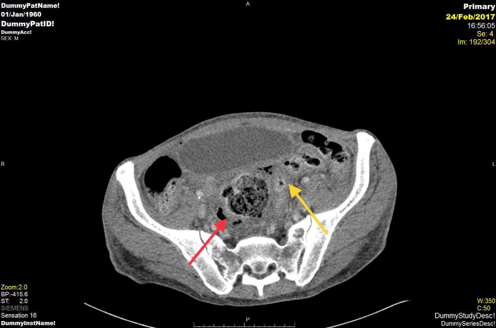
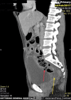
Figure 2:
Sagittal section of the CT image showing red arrow indicating extra luminal air close to sigmoid colon and Yellow arrow shows free fluid collection in keeping with the sigmoid perforation.
Read Full Article HTML DOI: 10.29328/journal.acst.1001009 Cite this Article Read Full Article PDF
More Images
Similar Articles
-
Theranostics: A Unique Concept to Nuclear MedicineLucio Mango*. Theranostics: A Unique Concept to Nuclear Medicine. . 2017 doi: 10.29328/journal.acst.1001001; 1: 001-004
-
Predictors of Candidemia infections and its associated risk of mortality among adult and pediatric cancer patients: A retrospective study in Lahore, Punjab, PakistanHafiz Muhammad Bilal*,Neelam Iqbal,Shazia Ayaz. Predictors of Candidemia infections and its associated risk of mortality among adult and pediatric cancer patients: A retrospective study in Lahore, Punjab, Pakistan. . 2018 doi: 10.29328/journal.acst.1001003; 2: 001-007
-
Endogenus toxicology: Modern physio-pathological aspects and relationship with new therapeutic strategies. An integrative discipline incorporating concepts from different research discipline like Biochemistry, Pharmacology and ToxicologyLuisetto M*,Naseer Almukhtar,Behzad Nili Ahmadabadi,Gamal Abdul Hamid,Ghulam Rasool Mashori,Kausar Rehman Khan,Farhan Ahmad Khan,Luca Cabianca. Endogenus toxicology: Modern physio-pathological aspects and relationship with new therapeutic strategies. An integrative discipline incorporating concepts from different research discipline like Biochemistry, Pharmacology and Toxicology . . 2019 doi: 10.29328/journal.acst.1001004; 3: 001-024
-
Insilico investigation of TNFSF10 signaling cascade in ovarian serous cystadenocarcinomaAsima Tayyeb*,Zafar Abbas Shah. Insilico investigation of TNFSF10 signaling cascade in ovarian serous cystadenocarcinoma. . 2019 doi: 10.29328/journal.acst.1001005; 3: 025-034
-
Fifth “dark” force completely change our understanding of the universeRobert Skopec*. Fifth “dark” force completely change our understanding of the universe. . 2019 doi: 10.29328/journal.acst.1001006; 3: 035-041
-
Risk factors of survival in breast cancerAkram Yazdani*. Risk factors of survival in breast cancer. . 2019 doi: 10.29328/journal.acst.1001007; 3: 042-044
-
Results of chemotherapy in the treatment of chronic lymphoid leukemia in Black Africa: Experience of Côte d’IvoirePacko Dieu-le-veut Saint-Cyr Sylvestre*,N’dhatz Comoe Emeraude,Kamara Ismael,Boidy Kouakou,Koffi Kouassi Gustave,Nanho Danho Clotaire,Koffi Kouassi Gustave. Results of chemotherapy in the treatment of chronic lymphoid leukemia in Black Africa: Experience of Côte d’Ivoire. . 2019 doi: 10.29328/journal.acst.1001008; 3: 045-048
-
Stercoral perforation: A rare case and reviewLava Krishna Kannappa*,Muhammad Sufian Khalid,May Hnin Lwin Ko,Mohsin Hussein,Jia Hui Choong,Ameer Omar Rawal-Pangarkar,Danaradja Armugam,Yahya Salama. Stercoral perforation: A rare case and review. . 2019 doi: 10.29328/journal.acst.1001009; 3: 049-051
-
Different optimization strategies for the optimal control of tumor growthAbd El Moniem NK*,Sweilam NH,Tharwat AA. Different optimization strategies for the optimal control of tumor growth. . 2019 doi: 10.29328/journal.acst.1001010; 3: 052-062
-
Risk factor of liver metastases in breast cancerAkram Yazdani*. Risk factor of liver metastases in breast cancer. . 2019 doi: 10.29328/journal.acst.1001011; 3: 063-065
Recently Viewed
-
Agriculture High-Quality Development and NutritionZhongsheng Guo*. Agriculture High-Quality Development and Nutrition. Arch Food Nutr Sci. 2024: doi: 10.29328/journal.afns.1001060; 8: 038-040
-
A Low-cost High-throughput Targeted Sequencing for the Accurate Detection of Respiratory Tract PathogenChangyan Ju, Chengbosen Zhou, Zhezhi Deng, Jingwei Gao, Weizhao Jiang, Hanbing Zeng, Haiwei Huang, Yongxiang Duan, David X Deng*. A Low-cost High-throughput Targeted Sequencing for the Accurate Detection of Respiratory Tract Pathogen. Int J Clin Virol. 2024: doi: 10.29328/journal.ijcv.1001056; 8: 001-007
-
A Comparative Study of Metoprolol and Amlodipine on Mortality, Disability and Complication in Acute StrokeJayantee Kalita*,Dhiraj Kumar,Nagendra B Gutti,Sandeep K Gupta,Anadi Mishra,Vivek Singh. A Comparative Study of Metoprolol and Amlodipine on Mortality, Disability and Complication in Acute Stroke. J Neurosci Neurol Disord. 2025: doi: 10.29328/journal.jnnd.1001108; 9: 039-045
-
Development of qualitative GC MS method for simultaneous identification of PM-CCM a modified illicit drugs preparation and its modern-day application in drug-facilitated crimesBhagat Singh*,Satish R Nailkar,Chetansen A Bhadkambekar,Suneel Prajapati,Sukhminder Kaur. Development of qualitative GC MS method for simultaneous identification of PM-CCM a modified illicit drugs preparation and its modern-day application in drug-facilitated crimes. J Forensic Sci Res. 2023: doi: 10.29328/journal.jfsr.1001043; 7: 004-010
-
A Gateway to Metal Resistance: Bacterial Response to Heavy Metal Toxicity in the Biological EnvironmentLoai Aljerf*,Nuha AlMasri. A Gateway to Metal Resistance: Bacterial Response to Heavy Metal Toxicity in the Biological Environment. Ann Adv Chem. 2018: doi: 10.29328/journal.aac.1001012; 2: 032-044
Most Viewed
-
Evaluation of Biostimulants Based on Recovered Protein Hydrolysates from Animal By-products as Plant Growth EnhancersH Pérez-Aguilar*, M Lacruz-Asaro, F Arán-Ais. Evaluation of Biostimulants Based on Recovered Protein Hydrolysates from Animal By-products as Plant Growth Enhancers. J Plant Sci Phytopathol. 2023 doi: 10.29328/journal.jpsp.1001104; 7: 042-047
-
Sinonasal Myxoma Extending into the Orbit in a 4-Year Old: A Case PresentationJulian A Purrinos*, Ramzi Younis. Sinonasal Myxoma Extending into the Orbit in a 4-Year Old: A Case Presentation. Arch Case Rep. 2024 doi: 10.29328/journal.acr.1001099; 8: 075-077
-
Feasibility study of magnetic sensing for detecting single-neuron action potentialsDenis Tonini,Kai Wu,Renata Saha,Jian-Ping Wang*. Feasibility study of magnetic sensing for detecting single-neuron action potentials. Ann Biomed Sci Eng. 2022 doi: 10.29328/journal.abse.1001018; 6: 019-029
-
Pediatric Dysgerminoma: Unveiling a Rare Ovarian TumorFaten Limaiem*, Khalil Saffar, Ahmed Halouani. Pediatric Dysgerminoma: Unveiling a Rare Ovarian Tumor. Arch Case Rep. 2024 doi: 10.29328/journal.acr.1001087; 8: 010-013
-
Physical activity can change the physiological and psychological circumstances during COVID-19 pandemic: A narrative reviewKhashayar Maroufi*. Physical activity can change the physiological and psychological circumstances during COVID-19 pandemic: A narrative review. J Sports Med Ther. 2021 doi: 10.29328/journal.jsmt.1001051; 6: 001-007

HSPI: We're glad you're here. Please click "create a new Query" if you are a new visitor to our website and need further information from us.
If you are already a member of our network and need to keep track of any developments regarding a question you have already submitted, click "take me to my Query."


